Fiber Microdriver
May 16, 2022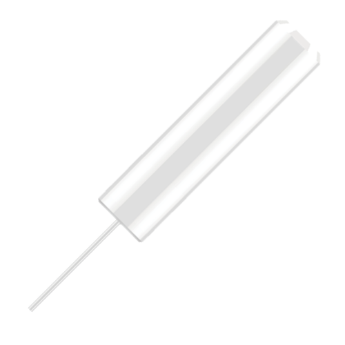

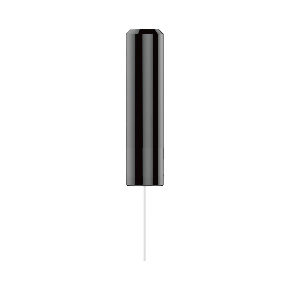
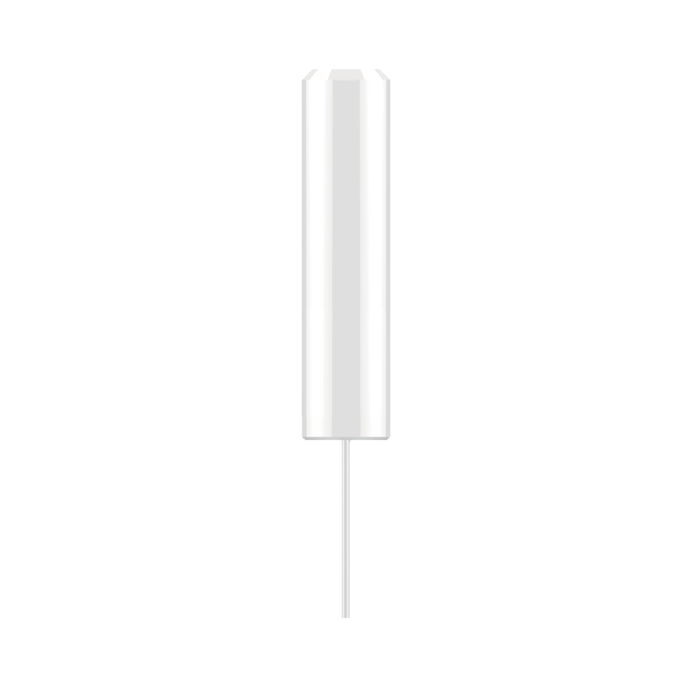

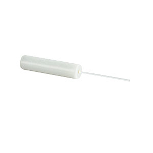
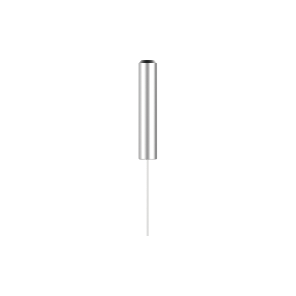
Fiber Optic Cannula
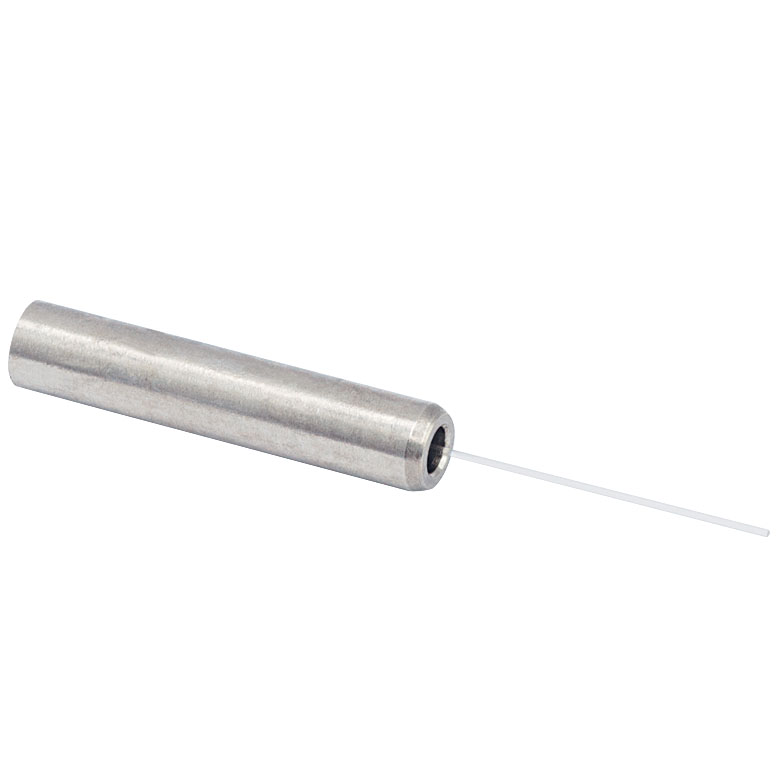
A single fiber-optic cannula consists of a cannula, called ferrule, and an optic fiber to guide the light from the light source to the brain target.
Original price was: € 55,50.€ 44,40Current price is: € 44,40.
Implantable Fiber Optic Cannulae transmit light to brain tissue from a light source for optogenetic manipulation or photometry recording purposes. The cannulae are made of a ferrule and an optical fiber which is cemented to the skull during a stereotaxic surgery. 50%-60% of the ferrule stays exposed after implanting, providing an interface to be connected to the fiber-optic patch cord to guide the light.
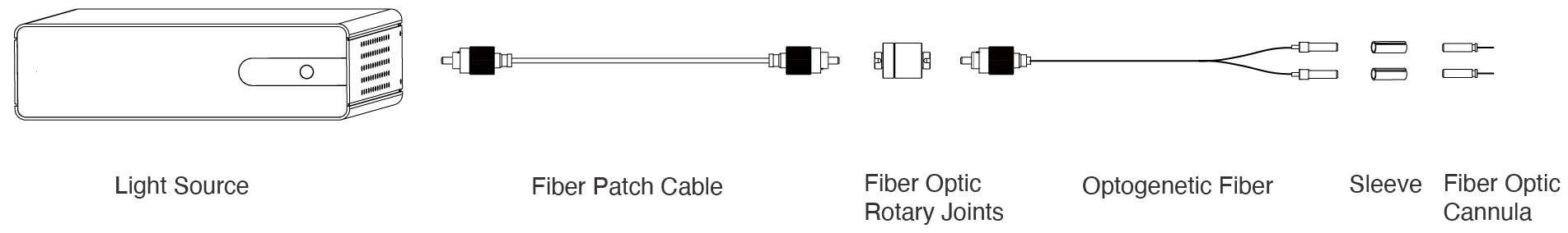
Implantation of the fiber optics into the cranium consists of:
- All surface of the cranium is exposed and connective tissue is cleared.
- The fiber optic implant is held in a situation with the stereotaxic arm.
- Dental cement is applied to fix the fiber optic implant to the skull. 50%-60% of the ferrule is exposed to be connected to the fiber-optic patch cord during
experiment.

| Weight | N/A |
|---|---|
| Dimensions | N/A |
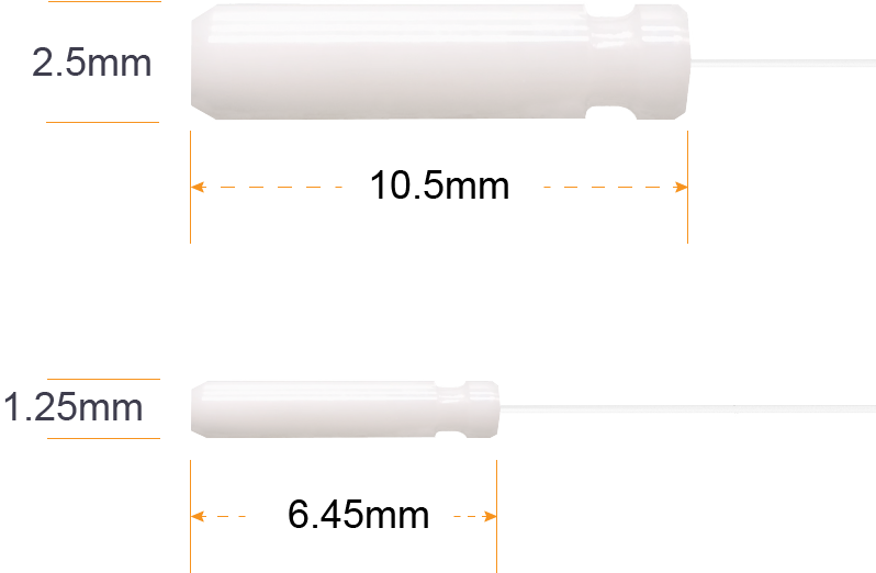
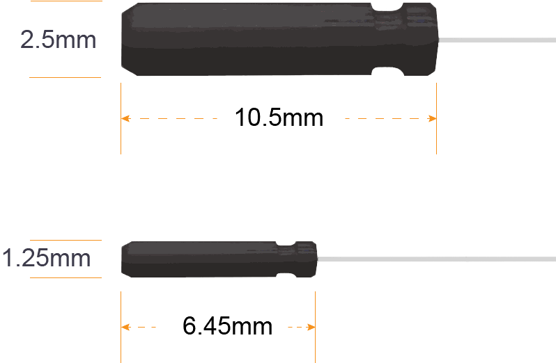
| Ferrule Material | Ceramic, Stainless Steel |
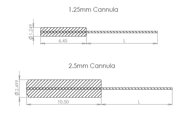
| Ferrule Outer Diameter | Ø1.25mm, Ø2.5mm |
| Ferrule Length | 10.5mm, 6.4mm |
| Fiber Diameter | Ø200µm, Ø400µm |
| Fiber Length | 10mm, 2mm, 5mm, Custom |
| Numerical Aperture (NA) | 0.22, 0.39, 0.50 |
Reviews
There are no reviews yet.